Featured post

Biology cell mcq

1. Who discovered cell in 1665? a) Robert Hook b) Robert Crook c) David Thomson d) Marie Francois Ans. a) 2. Name an Organelle ...

Friday, 20 December 2019

Structure and Function of the Eyes


.
The structures and functions of the eyes are complex. Each eye constantly adjusts the amount of light it lets in, focuses on objects near and far, and produces continuous images that are instantly transmitted to the brain.
The orbit is the bony cavity that contains the eyeball, muscles, nerves, and blood vessels, as well as the structures that produce and drain tears. Each orbit is a pear-shaped structure that is formed by several bones. 
आंखों की संरचना और कार्य जटिल हैं।  प्रत्येक आंख लगातार प्रकाश की मात्रा को समायोजित करती है जो इसे अंदर जाने देती है, निकट और दूर की वस्तुओं पर ध्यान केंद्रित करती है, और निरंतर छवियों का उत्पादन करती है जो तुरंत मस्तिष्क में प्रेषित होती हैं।

 कक्षा हड्डी गुहा है जिसमें नेत्रगोलक, मांसपेशियां, तंत्रिकाएं और रक्त वाहिकाएं होती हैं, साथ ही साथ संरचनाएं भी होती हैं जो आँसू पैदा करती हैं और निकलती हैं।  प्रत्येक कक्षा एक नाशपाती के आकार की संरचना है जो कई हड्डियों द्वारा बनाई जाती है।

Key Points

  • The eye is made up of a number of parts, including the iris, pupil, cornea, and retina.
  • The eye has six muscles which control the eye movement, all providing different tension and torque.
  • The eye works a lot like a camera, the pupil provides the f-stop, the iris the aperture stop, the cornea resembles a lens. The way that the image is formed is much like the way a convex lens forms an image.

Key Terms


  • pupil: The hole in the middle of the iris of the eye, through which light passes to be focused on the retina.
  • aperture: The diameter of the aperture that restricts the width of the light path through the whole system. For a telescope, this is the diameter of the objective lens (e.g., a telescope may have a 100 cm aperture).


प्रमुख बिंदु

 आंख कई हिस्सों से बनी है, जिसमें आईरिस, पुतली, कॉर्निया और रेटिना शामिल हैं।

 आंख में छह मांसपेशियां होती हैं जो आंखों की गति को नियंत्रित करती हैं, सभी अलग-अलग तनाव और टॉर्क प्रदान करती हैं।

 आंख एक कैमरे की तरह बहुत काम करती है, पुतली एफ-स्टॉप प्रदान करती है, आईरिस एपर्चर स्टॉप, कॉर्निया एक लेंस जैसा दिखता है।  जिस तरह से छवि का निर्माण होता है, वह उस तरह से होता है, जिस तरह से उत्तल लेंस एक छवि बनाता है।

 मुख्य शर्तें

 पुतली: आंख के परितारिका के बीच का छेद, जिससे होकर प्रकाश रेटिना पर केंद्रित होता है।

 एपर्चर: एपर्चर का व्यास जो पूरे सिस्टम के माध्यम से प्रकाश पथ की चौड़ाई को प्रतिबंधित करता है।  टेलीस्कोप के लिए, यह ऑब्जेक्टिव लेंस का व्यास है (उदाहरण के लिए, टेलीस्कोप में 100 सेमी एपर्चर हो सकता है)।

The human eye is the gateway to one of our five senses. The human eye is an organ that reacts with light. It allows light perception, color vision and depth perception. A normal human eye can see about 10 million different colors! There are many parts of a human eye, and that is what we are going to cover in this atom.

Properties

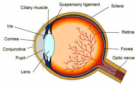
Contrary to what you might think, the human eye is not a perfect sphere, but is made up of two differently shaped pieces, the cornea and the sclera. These two parts are connected by a ring called the limbus. The part of the eye that is seen is the iris, which is the colorful part of the eye. In the middle of the iris is the pupil, the black dot that changes size. The cornea covers these elements, but is transparent. The fundus is on the opposite of the pupil, but inside the eye and can not be seen without special instruments. The optic nerve is what conveys the signals of the eye to the brain. is a diagram of the eye. The human eye is made up of three coats:
  1. Outermost Layer – composed of the cornea and the sclera.
  2. Middle Layer – composed of the choroid, ciliary body and iris.
  3. Innermost Layer – the retina, which can be seen with an instrument called the ophthalmoscope.
Once you are inside these three layers, there is the aqueous humor (clear fluid that is contained in the anterior chamber and posterior chamber), vitreous body (clear jelly that is much bigger than the aqueous humor), and the flexible lens. All of these are connected by the pupil.
मानव आंख हमारी पांच इंद्रियों में से एक का प्रवेश द्वार है।  मानव आँख एक अंग है जो प्रकाश के साथ प्रतिक्रिया करता है।  यह प्रकाश धारणा, रंग दृष्टि और गहराई की धारणा की अनुमति देता है।  एक सामान्य मानव आँख लगभग 10 मिलियन विभिन्न रंगों को देख सकती है!  एक इंसानी आंख के कई हिस्से हैं, और यही हम इस परमाणु में शामिल होने जा रहे हैं।

 गुण

 आप जो सोच सकते हैं, उसके विपरीत, मानव आंख एक संपूर्ण क्षेत्र नहीं है, लेकिन दो अलग-अलग आकार के टुकड़ों, कॉर्निया और श्वेतपटल से बना है।  ये दोनों भाग एक रिंग से जुड़े होते हैं जिसे लिमबस कहा जाता है।  आंख का जो हिस्सा दिखाई पड़ता है वह परितारिका है, जो आंख का रंगीन हिस्सा है।  परितारिका के बीच में पुतली है, काली बिंदी जो आकार बदलती है।  कॉर्निया इन तत्वों को कवर करता है, लेकिन पारदर्शी है।  निधि पुतली के विपरीत पर है, लेकिन आंख के अंदर और विशेष उपकरणों के बिना नहीं देखा जा सकता है।  ऑप्टिक तंत्रिका वह है जो आंख के संकेतों को मस्तिष्क तक पहुंचाती है।  आंख का एक आरेख है।  मानव आँख तीन कोटों से बनी होती है:
बाह्य परत - कॉर्निया और श्वेतपटल से बना है।

 मध्य परत - कोरॉइड, सिलिअरी बॉडी और आइरिस से बना है।

 इनरमोस्ट लेयर - रेटिना, जिसे ओफ्थाल्मोस्कोप नामक उपकरण के साथ देखा जा सकता है।

 एक बार जब आप इन तीन परतों के अंदर होते हैं, तो जलीय हास्य होता है (स्पष्ट तरल पदार्थ जो पूर्वकाल कक्ष और पीछे के कक्ष में निहित होता है), विट्रीस बॉडी (स्पष्ट जेली जो जलीय हास्य की तुलना में बहुत बड़ा होता है), और लचीला लेंस होता है।  ये सभी पुतली द्वारा जुड़े हुए हैं।
:
image
Diagram of the Human Eye: The cornea and lens of an eye act together to form a real image on the light-sensing retina, which has its densest concentration of receptors in the fovea and a blind spot over the optic nerve. The power of the lens of an eye is adjustable to provide an image on the retina for varying object distances. Layers of tissues with varying indices of refraction in the lens are shown here. However, they have been omitted from other pictures for clarity.

Thursday, 19 December 2019

Leukemia


Leukemia is a cancer of the blood or bone marrow. Bone marrow produces blood cells. Leukemia can develop due to a problem with blood cell production. It usually affects the leukocytes, or white blood cells.
Leukemia is most likely to affect people over the age of 55 years, but it is also the most common cancer in those aged under 15 years.
The National Cancer Institute estimates that 61,780 people will receive a diagnosis of leukemia in 2019. They also predict that leukemia will cause 22,840 deaths in the same year.
Acute leukemia develops quickly and worsens rapidly, but chronic leukemia gets worse over time. There are several different types of leukemia, and the best course of treatment and a person's chance of survival depends on which type they have.
Leukemia develops when the DNA of developing blood cells, mainly white cells, incurs damage. This causes the blood cells to grow and divide uncontrollably.
Healthy blood cells die, and new cells replace them. These develop in the bone marrow.
The abnormal blood cells do not die at a natural point in their life cycle. Instead, they build up and occupy more space.
As the bone marrow produces more cancer cells, they begin to overcrowd the blood, preventing the healthy white blood cells from growing and functioning normally.
Eventually, the cancerous cells outnumber healthy cells in the blood.

There is a range of risk factors for leukemia. Some of these risk factors have more significant links to leukemia than others:
Artificial ionizing radiation: This could include having received radiation therapy for a previous cancer, although this is a more significant risk factor for some types than others.
Certain viruses: The human T-lymphotropic virus (HTLV-1) has links to leukemia.
Chemotherapy: People who received chemotherapy treatment for a previous cancer have a higher chance of developing leukemia later in life.
Exposure to benzene: This is a solvent that manufacturers use in some cleaning chemicals and hair dyes.
Some genetic conditions: Children with Down syndrome have a third copy of chromosome 21. This increases their risk of acute myeloid or acute lymphocytic leukemia to 2–3%, which is higher than in children without this syndrome.
Another genetic condition with links to leukemia is Li-Fraumeni syndrome. This causes a change to the TP53 gene.
Family history: Having siblings with leukemia can lead to a low but significant risk of leukemia. If a person has an identical twin with leukemia, they have a 1 in 5 chance of having the cancer themselves.
Inherited problems with the immune system: Certain inherited immune conditions increase the risk of both severe infections and leukemia. These include:
  • ataxia-telangiectasia
  • Bloom syndrome
  • Schwachman-Diamond syndrome
  • Wiskott-Aldrich syndrome
Immune suppression: Childhood leukemia may develop due to the deliberate suppression of the immune system. This might occur following an organ transplant as a child takes medications to prevent their body from rejecting the organ.
Several risk factors need further studies to confirm their link to leukemia, such as:
  • exposure to electromagnetic fields
  • exposure to certain chemicals in the workplace, such as gasoline, diesel, and pesticides
  • smoking
  • using hair dyes

There are four main categories of leukemia:
  • acute
  • chronic
  • lymphocytic
  • myelogenous
Chronic and acute leukemias
During its lifespan, a white blood cell goes through several stages.
In acute leukemia, developing cells multiply quickly and collect in the marrow and blood. They exit the bone marrow too early and are not functional.
Chronic leukemia progresses more slowly. It allows for the production of more mature, useful cells.
Acute leukemia overcrowds the healthy blood cells more quickly than chronic leukemia.
Lymphocytic and myelogenous leukemias
Doctors classify leukemia according to the type of blood cell they affect.
Lymphocytic leukemia occurs if cancerous changes affect the type of bone marrow that makes lymphocytes. A lymphocyte is a white blood cell that plays a role in the immune system.
Myelogenous leukemia happens when the changes affect bone marrow cells that produce blood cells, rather than the blood cells themselves.

Acute lymphocytic leukemia

Children under 5 years old are at the highest risk of developing acute lymphoblastic leukemia (ALL). However, It can also affect adults, typically over the age of 50 years. Out of every five deaths from ALL, four occur in adults.

Chronic lymphocytic leukemia

This is most common among adults over 55 years, but younger adults can also develop it. About 25% of adults with leukemia have chronic lymphocytic leukemia (CLL). It is more common in men than in women and rarely affects children.

Acute myelogenous leukemia

Acute myelogenous leukemia (AML) is more common in adults than in children, but overall, it is a rare cancer. It develops more often in men than in women.
It develops quickly, and symptoms include fever, difficulty breathing, and pain in the joints. Environmental factors can trigger this type.

Chronic myeloid leukemia

Chronic myelogenous leukemia (CML) mostly develops in adults. About 15% of all leukemia cases in the United States are CML. Children rarely develop this type of leukemia.

Monday, 16 December 2019

Nervous System

The Nervous System is a very important part of the animal body that coordinates voluntary and involuntary actions of the body.

What is Nerve Impulse?

Any stimulus or signal that activate or inhibit any gland, muscle, organ, tissue or any nerve cell as known as Nerve impulse. For Example: When we touch a hot plate, a signal is transmitted to brain due to which we respond by removing our hand.
There are two  types of Nervous System in vertebrates:vertebrates:
  • Central Nervous System
  • Peripheral Nervous System
Divisions of Nervous System
Fig.1. Divisions of Nervous System

What is Central Nervous System?

The central Nervous System comprises of Brainand the Spinal Cord.  

Explain the Structure of Human Brain.

The Human Brain is covered by a membrane known as Meninges. Meninges are three in number. The Outermost DuramaterMiddle Arachnoid and Innermost Piamater.
The longitudinal fissure divides the brain into two hemispheres known as Right Cerebral Hemisphere and Left Cerebral Hemisphere.
The Brain is divided into three parts: ForebrainMidbrain and Hindbrain.  

Forebrain

The Forebrain comprises of Cerebrum,Thalamusand Hypothalamus.
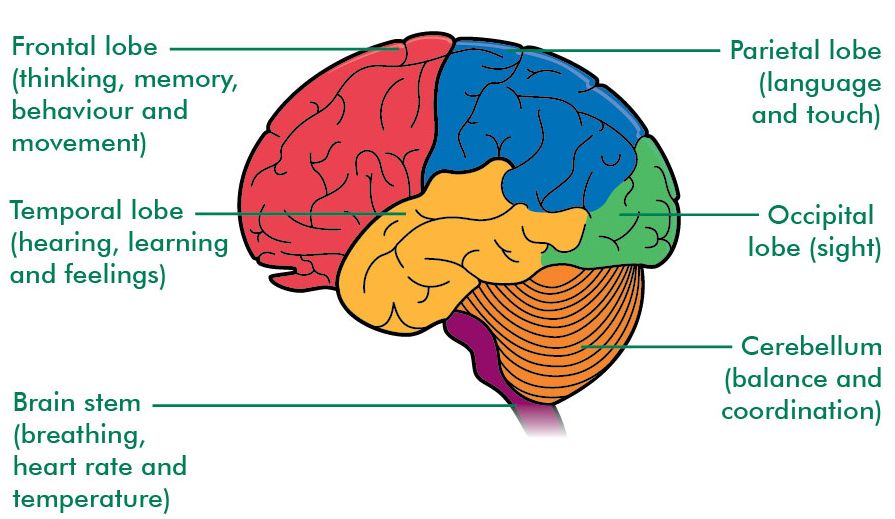
  • Cerebrum is the largest part of the human brain. It is divided into four lobes: Occipital LobeParietal LobeTemporal Lobe and Frontal Lobe.
Different Lobes of Cerebrum and their function
Fig.2. Different Lobes of Cerebrum and their function
Thalamus is olive shaped structure which performs sensory functions. It directs sensory input from the eyes, ears, tongue and from other sense organs.
Hypothalamus is located below the thalamus as the name suggests. It is important in monitoring hormone concentrations, water concentrations, temperature of the body etc.  

Midbrain

It is a small part of the brain. It is associated with Vison, Hearing, Sleep, Arousal, Temperature etc.  

Hindbrain

It consists of Cerebellum, Pons and Medulla Oblongata.
  • Cerebellum is involved in controlling attention, language, equilibrium, posture, etc.
  • Pons controls digestive and respiratory movements.
  • Medulla Oblongata connects skull with the spinal cord. It helps in controlling Blood Pressure, Heart Rate, Respiratory Movement, Vomiting etc.  

What are Neurons/Nerve Cell?

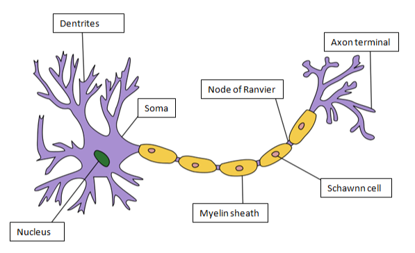
The structural and the functional unit of brain is NeuronNeuron is divided into Cell BodyAxonand Dendrites. Cell body/soma of the neuron contain nucleus. It also contains Nissl Granules, composed of endoplasmic reticulum. Dendrites are branched projections from the cell body. Axon is a long, slender in shape that transmits nerve impulse from one neuron to another. 
Structure of Neuron
Fig.3. Structure of Neuron
The cells found in axon are known as Schawnn Cell. Myelin sheath is schawnncells. Myelin sheath is an electrically insulating layer which is required for fast and efficient nerve impulse transmission. A gap between adjacent Schawnn Cells is known as Nodes of Ranvier.

Example of different types of Neurons

  • Sensory Neurons transmit nerve impulses from sensory receptors to central nervous system.
  • Motor Neurons transmit nerve impulses from central nervous system to muscle or a gland.
  • Inter Neurons connects different neurons within the brain and spinal cord.

Spinal Nerves Spinal Cord

It is a thin, tube-like structure which extends from the medulla of the brain to the lumbar region of vertebral column. It helps in transmission of nerve impulses from the brain to the rest of the body. It also coordinates reflex action.
There are 31 pairs of Spinal Nerves on each side of the vertebral column. These Spinal Nerves are grouped into:
  • 8 pairs of cervical nerves
  • 12 pairs of thoracic nerves
  • 5 pairs of lumbar nerves
  • 5 pairs of sacral nerves
  • One pair of coccygeal nerves
The Spinal Cord is made up of White Matter and Gray Matter. The butterfly shaped structure present at the center of the spinal cord is known as Gray Matter
Structure of Spinal Cord
Fig.5. Structure of Spinal Cord
The region around the gray matter is known as White Matter. White matter contains nerves that are covered with Myelin Sheath whereas gray matter contains nerves without myelin sheath.  
What is Reflex Action?
The involuntary response towards any stimulus is known as Reflex Action.
What is Reflex Arc?
The pathway of reflex action is known as Reflex Arc. Some sensory neurons do not pass directly into the brain; they innervate in the spinal cord. This reduces the time of reflex action.
What is Peripheral Nervous System?
It consists of nerves and ganglia. Ganglia is a cluster of nerves. Peripheral Nervous System connects central nervous system to different organs and limbs.
Peripheral nervous system is divided into Autonomic Nervous System and Somatic Nervous System.  Autonomic nervous system is associated with communication to internal organs and glands. Somatic nervous system communicates with sensory organs and voluntary muscles.
Autonomic Nervous System is further divided into Parasympathetic Nervous System and Sympathetic Nervous System.

Difference between Sympathetic and Parasympathetic Nervous System

Sympathetic Nervous System
Parasympathetic Nervous System
Type of Control
Involuntary
Involuntary
Number of neurons per message
Two (Preganglionic shorter than Postganglionic)
Two (Preganglionic longer than Postganglionic)
Location of motor fiber
Thoracolumbar spinal nerves
Cranial (e.g., vagus) and sacral spinal nerves
Neurotransmitter
Norepinephrine
Acetylcholine
Effectors
Smooth and cardiac muscle, glands

Human Sense Organs

Humans have five sense organs, They are: Eyes, Ears, Nose, Skin and Tongue.
The structure of Eye and Ear is explained below:

Structure of Human Eye

Structure of the Eye

Eye is a sense organ for sight. It consists of three layers: Outermost Sclera, Middle Choroid which is supplied with blood vessels and Inner Retina. The membrane that covers the sclera is known as Conjunctiva. Retina contains light sensitive cells known as Rods and Cones. Rods are cells that allow vision in dim light whereas cones help in color vision. Light enters the eye via cornea. 
The nerve that connect eye to brain is known as Optic Nerve. Transparent lens focuses light on retina which helps in vision. The muscles that contracts and relaxes the lens is known as Suspensory Ligament. This is dependent on the amount of light that falls on the lens.
The colored part of the eye that controls the amount of light that enters the eye is known as Pupil. Fovea is an exact location where light is focused on the retina.   

Structure of Ear

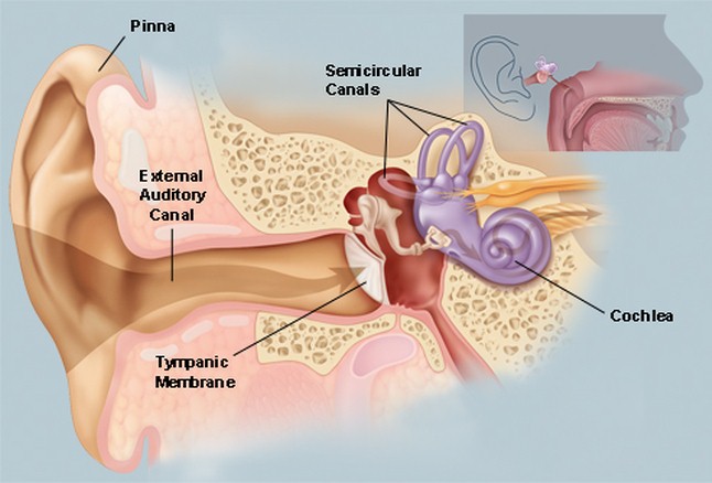
Ear is meant for hearing. The ear is divided into External EarMiddle Ear and Inner Ear. The outer part of the ear is known as Ear Pinna. Ear pinna is covered by a membrane known as Tympanic Membrane
Structure of the Ear
Fig. 7. Structure of the Ear
When sound strikes the pinna it vibrates and vibrations are passed to cochlea which is a part of the inner ear. There are fluid-filled canals present that are attached to cochlea, are known as Semi-Circular Canals. These canals help in maintaining equilibrium or balance of the body.

Stimulus and Response

It is defined as any detectable change in the internal or the external environment. The ability of an organism to react against the stimulus is known as Response.

Synapse

The junction between the two Nerve cells is known as Synapse.  

Second Brain in Humans

The Enteric Nervous System is known as Second Brain in Humans.  

Saltatory Conduction

During Nerve Impulse transmission, the nerve impulse jumps over the Nodes of Ranvier. This mode of nerve impulse transmission is known as Salutatory Conduction. This is the fast mode of nerve impulse transmission.